Key takeaways
- This page offers 25 free RCIS practice questions with fully worked explanations, requiring no signup or payment.
- The questions are grouped into six blueprint-matched topics: vascular access and angiographic technique, diagnostic catheterization and hemodynamics, PCI and intracoronary imaging, structural interventions and mechanical support, pharmacology and sedation, and emergencies and radiation safety.
- These are original questions written to the CCI RCIS content blueprint, not reproductions of real exam items.
- The Cardiac Codex app's full practice bank contains 2,198 questions with explanations, plus two full-length, live and timed RCIS mock exams.
- A free 25-question RCIS diagnostic that scores results by topic and recommends where to focus is also available.
Vascular access & angiographic technique
-
1What is the recommended anatomical landmark for initial puncture during transradial access to avoid the flexor retinaculum?
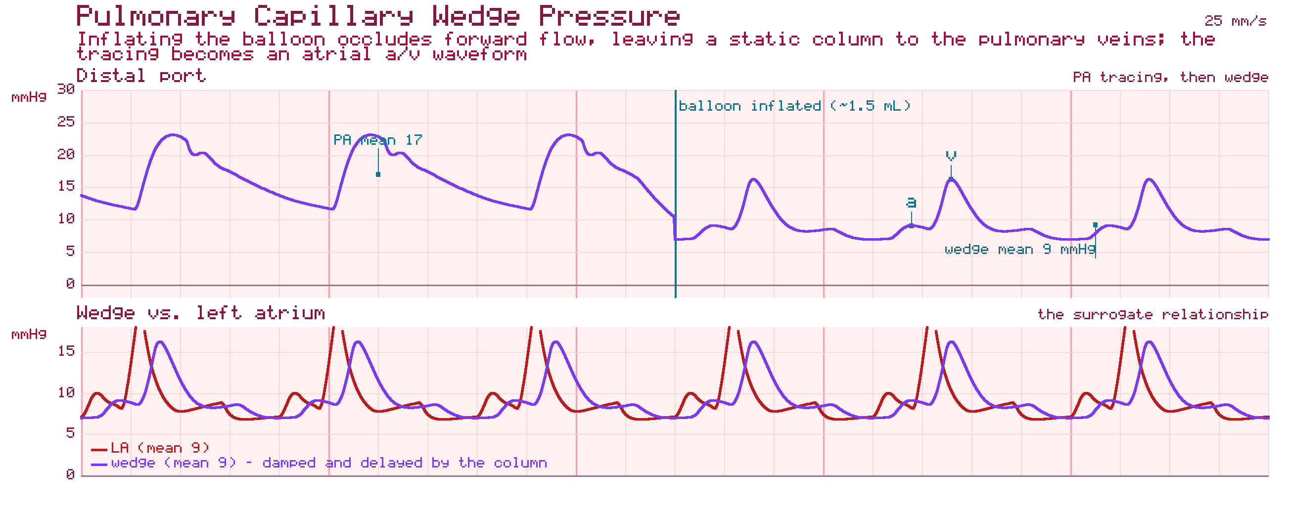
- A5 cm distal to the radial styloid process
- BAt the radial styloid process
- C2 cm proximal to the radial styloid process
- DAt the level of the antecubital fossa
Correct answer: C — 2 cm proximal to the radial styloid process
Accessing 2 cm proximal to the radial styloid process avoids the flexor retinaculum and the tapering branches of the artery, whereas other sites increase risk of structural complications.
-
2Current clinical guidelines strongly endorse a 'radial-first' approach for diagnostic angiography and PCI primarily because of which benefit?
- ALower contrast volume requirements
- BFaster overall procedure completion in every case
- CElimination of the need for anticoagulation
- DProfound reductions in access-site bleeding, major vascular complications, and mortality
Correct answer: D — Profound reductions in access-site bleeding, major vascular complications, and mortality
The radial-first paradigm shift is driven by profound reductions in access-site bleeding, major vascular complications, and overall mortality, particularly in ACS patients.
-
3Entering the aortic arch from a right radial approach typically requires which catheter curve, compared with the JL4.0 standard used femorally?
- AA larger curve, such as a JL5.0
- BThe identical JL4.0 curve
- CA smaller curve, such as a JL3.5
- DA pigtail catheter instead of a Judkins curve
Correct answer: C — A smaller curve, such as a JL3.5
Entering the aortic arch from the right radial approach requires a smaller catheter curve than femoral access, such as a JL3.5 instead of a JL4.0, to achieve stable seating.
-
4The right coronary artery originates from which aortic sinus?
- AThe left coronary sinus
- BThe right posterior (non-coronary) sinus
- CThe anterior (right) coronary sinus
- DThe commissure between the left and non-coronary sinuses
Correct answer: C — The anterior (right) coronary sinus
The RCA originates from the anterior coronary sinus, while the LMCA originates from the left coronary sinus.
-
5While positioning for a coronary angiogram, the operator selects a right anterior oblique (RAO) view. To confirm correct orientation, the thoracic spine should appear on which side of the image?
- AThe right
- BThe center
- CIt alternates depending on cranial or caudal angulation
- DThe left
Correct answer: D — The left
In the RAO view, the image intensifier is on the patient's right, so the thoracic spine appears on the left of the screen with ribs descending to the right.
Diagnostic catheterization & hemodynamics
-
6Despite the availability of cardiac MRI, CT angiography, and advanced echocardiography, invasive cardiac catheterization remains the gold standard primarily because of which unique capability?
- AIt is the only modality that can visualize the coronary arteries at all
- BIt carries zero procedural risk
- CIt requires no contrast media
- DIt precisely quantifies pressure gradients and directly informs interventional or surgical planning
Correct answer: D — It precisely quantifies pressure gradients and directly informs interventional or surgical planning
Despite advances in non-invasive imaging, catheterization remains the gold standard because it uniquely and precisely quantifies pressure gradients, resolves ambiguous clinical presentations, and directly informs interventional or surgical planning.
-
7A complete blood count (CBC) is obtained before catheterization primarily to assess which risk?
- AContrast-induced nephropathy risk
- BHemorrhagic complication risk, via baseline hemoglobin and platelet levels
- CArrhythmia risk
- DRadial artery spasm risk
Correct answer: B — Hemorrhagic complication risk, via baseline hemoglobin and platelet levels
A CBC establishes baseline hemoglobin and platelet levels, quantifying the risk of hemorrhagic complications given the anticipated use of high-dose anticoagulant and antiplatelet medications.
-
8Left ventricular end-diastolic pressure (LVEDP) normally ranges between:
- A4 to 12 mmHg
- B25 to 35 mmHg
- C0 to 5 mmHg
- D15 to 25 mmHg
Correct answer: A — 4 to 12 mmHg
In a healthy adult, resting LVEDP typically falls within 4 and 12 mmHg, with higher values indicating impaired relaxation or overt diastolic dysfunction.
-
9Which of the following is a classic clinical feature of hypovolemic shock?
- ANormal filling pressures and normal SVR
- BLow PCWP and high Systemic Vascular Resistance
- CLow SVR and elevated Central Venous Pressure
- DHigh PCWP and high Cardiac Output
Correct answer: B — Low PCWP and high Systemic Vascular Resistance
Hypovolemic shock is characterized by a reduction in filling pressures (low PCWP) and high SVR due to compensatory vasoconstriction.
-
10Which intracoronary imaging modality provides microscopic, cross-sectional imaging of the vessel wall to characterize plaque composition and ensure precise stent expansion?
- AOptical Coherence Tomography (OCT)
- BFractional Flow Reserve (FFR)
- CTransesophageal Echocardiography (TOE)
- DFluoroscopy
Correct answer: A — Optical Coherence Tomography (OCT)
Both OCT and Intravascular Ultrasound (IVUS) provide high-resolution cross-sectional images of the coronary vessel wall to optimize stent apposition.
PCI & intracoronary imaging
-
11The fundamental mechanics of PCI involve gaining arterial access, advancing a guidewire across a stenotic lesion, and utilizing balloon dilation, followed by which final step?
- ADeployment of a metallic scaffold to prevent elastic recoil
- BImmediate surgical closure of the arteriotomy
- CInjection of a chemical sclerosing agent
- DPlacement of a temporary external compression device only
Correct answer: A — Deployment of a metallic scaffold to prevent elastic recoil
PCI mechanics conclude with deployment of a metallic scaffold (stent) to prevent elastic recoil after balloon dilation mechanically disrupts the plaque and stretches the arterial wall.
-
12IVUS utilizes high-frequency sound waves in what general range, and does NOT require which additional step during acquisition?
- A1-2 MHz, and requires contrast clearance in every case
- B300-400 MHz, and requires general anesthesia
- C40-60 MHz, and does not require contrast clearance
- D40-60 MHz, but requires a full week of preparation
Correct answer: C — 40-60 MHz, and does not require contrast clearance
IVUS uses 40-60 MHz sound waves, which easily penetrate the blood pool, meaning image acquisition does not require contrast clearance unlike OCT.
-
13Which trial found that OCT-guided PCI achieved a minimum stent area non-inferior to IVUS guidance, but failed to demonstrate superiority over angiography-guided PCI?
- AVALIDATE-SWEDEHEART
- BILUMIEN III: OPTIMIZE PCI
- CULTIMATE
- DPARTNER 3
Correct answer: B — ILUMIEN III: OPTIMIZE PCI
ILUMIEN III: OPTIMIZE PCI (Lancet, 2016) randomised 450 patients to OCT-, IVUS-, or angiography-guided PCI. Median minimum stent area was 5.79 mm² with OCT, 5.89 mm² with IVUS, and 5.49 mm² with angiography. OCT met non-inferiority versus IVUS (p=0.001) but was not superior to IVUS (p=0.42), and was not superior to angiography (p=0.12) — the hierarchical superiority tests failed.
-
14During an intervention on the right coronary artery, the patient develops profound sinus bradycardia, hypotension, and vasodilation without reflex tachycardia. What is this physiologic triad known as?
- ABezold-Jarisch Reflex
- BBainbridge Reflex
- CVasovagal Syncope
- DCushing's Triad
Correct answer: A — Bezold-Jarisch Reflex
The Bezold-Jarisch reflex is triggered by ischemia or stretch in the inferoposterior wall and manifests as bradycardia, hypotension, and vasodilation, distinctly lacking the reflex tachycardia seen in typical shock.
Structural interventions & mechanical support
-
15An intra-aortic balloon pump augments coronary and cerebral perfusion through which timing mechanism?
- ARemaining continuously inflated throughout the cardiac cycle
- BInflating during systole and deflating during diastole
- CInflating during early diastole and deflating just prior to systole
- DInflating only in response to a sensed arrhythmia
Correct answer: C — Inflating during early diastole and deflating just prior to systole
The IABP inflates during early diastole to augment diastolic aortic root pressure and coronary/cerebral perfusion, then rapidly deflates just prior to systole to reduce left ventricular afterload.
-
16The modern cardiac catheterization laboratory has evolved from managing epicardial coronary artery disease alone into a theater for which broader scope of practice?
- AStructural heart interventions, advanced tissue diagnostics, and hemodynamic support
- BExclusively diagnostic coronary angiography with no therapeutic capability
- COpen surgical valve replacement only
- DNon-invasive imaging exclusively
Correct answer: A — Structural heart interventions, advanced tissue diagnostics, and hemodynamic support
The cath lab has evolved from purely coronary disease management via balloon angioplasty and bare-metal stenting into a complex theater for structural heart interventions, tissue diagnostics, and hemodynamic support.
-
17TAVR complications are classified into which three primary categories?
- ADevice-related, vascular access-site, and systemic complications
- BOnly anesthesia-related complications
- COnly radiation-related complications
- DOnly contrast-related complications
Correct answer: A — Device-related, vascular access-site, and systemic complications
TAVR complications are classified into three primary categories: device-related (PVL, thrombosis), vascular access-site (pseudoaneurysm, dissection, AV fistula), and systemic (atheroembolism, stroke).
-
18Placement of multiple TEER clips on the mitral valve carries which specific procedural risk that must be monitored via continuous transmitral gradient measurement?
- AImmediate cardiac arrest in all cases
- BGuaranteed device embolization
- CIatrogenic mitral stenosis
- DPermanent AV block
Correct answer: C — Iatrogenic mitral stenosis
Transmitral gradients must be monitored continuously during multi-clip TEER procedures, as placing multiple clips risks creating iatrogenic mitral stenosis.
Pharmacology & sedation
-
19Unfractionated heparin exerts its anticoagulant effect primarily by:
- ABlocking the GPIIb/IIIa receptor on platelets
- BInhibiting vitamin K epoxide reductase
- CDirectly inhibiting thrombin without a cofactor
- DBinding antithrombin III and accelerating inhibition of thrombin and Factor Xa
Correct answer: D — Binding antithrombin III and accelerating inhibition of thrombin and Factor Xa
Heparin binds antithrombin III, greatly accelerating its natural inhibition of thrombin (IIa) and Factor Xa.
-
20The Ramsay Sedation Scale and the Richmond Agitation-Sedation Scale (RASS) serve which function during procedural sedation?
- AMeasuring the patient's coronary artery calcium score
- BAllowing consistent titration and documentation of sedation depth across staff
- CGrading the severity of aortic stenosis
- DClassifying coronary artery perforation
Correct answer: B — Allowing consistent titration and documentation of sedation depth across staff
The Ramsay Sedation Scale and RASS are standardized scales that allow consistent titration and documentation of sedation depth across different staff members.
-
21Unfractionated heparin (UFH) anticoagulation during PCI is monitored via which laboratory parameter?
- AInternational Normalized Ratio (INR)
- BActivated Clotting Time (ACT)
- CPlatelet count alone
- DSerum creatinine
Correct answer: B — Activated Clotting Time (ACT)
UFH remains the standard anticoagulant during PCI, monitored via Activated Clotting Time (ACT) to ensure appropriate anticoagulation intensity.
Emergencies & radiation safety
-
22A vasovagal reaction during sheath removal classically presents with which combination of findings?
- ATachycardia, hypotension, and fever
- BTachycardia, hypertension, and pallor
- CBradycardia, hypotension, and diaphoresis
- DBradycardia, hypertension, and flushing
Correct answer: C — Bradycardia, hypotension, and diaphoresis
Vasovagal reactions classically present with bradycardia, hypotension, pallor, and diaphoresis, often triggered by sheath manipulation, pain, or anxiety.
-
23What is the first step in the stepwise response to suspected cardiac perforation/tamponade?
- AAdminister a GPIIb/IIIa inhibitor
- BImmediately call cardiac surgery and wait for their arrival before doing anything else
- CIncrease the heparin infusion rate
- DNotify the operator and team immediately
Correct answer: D — Notify the operator and team immediately
Immediate notification of the operator and team is the essential first step, enabling rapid coordinated response.
-
24In radiation safety, the acronym ALARA directs staff to keep exposure at what level relative to clinical necessity?
- AAs Low As Reasonably Achievable
- BAs Long As Radiographically Adequate
- CAlways Limit All Radiographic Anomalies
- DAs Little Additional Radiation as Available
Correct answer: A — As Low As Reasonably Achievable
ALARA (As Low As Reasonably Achievable) is the guiding principle that every radiation exposure should be justified by clinical benefit and minimized without compromising the case's diagnostic or therapeutic goal.
-
25In the fluoroscopic imaging chain, which component produces the X-ray beam?
- AThe flat-panel detector
- BThe X-ray generator
- CThe C-arm mechanical gantry alone
- DThe dosimetry badge
Correct answer: B — The X-ray generator
The X-ray generator produces the beam, which passes through the patient and is captured by the detector, with the image displayed in real time.
Bonus questions · week of Oct 12, 2026
-
26What is the primary risk of puncturing the common femoral artery above the inguinal ligament?
- AAV fistula formation
- BRadial artery spasm
- CPseudoaneurysm formation
- DRetroperitoneal hemorrhage
Correct answer: D — Retroperitoneal hemorrhage
Puncturing above the inguinal ligament enters the external iliac artery, which is outside the compressible area of the femoral head, leading to retroperitoneal hemorrhage.
-
27A patient has a normal ejection fraction on echocardiography but exertional dyspnea. Invasive hemodynamics reveal elevated filling pressures. Which diagnosis is this pattern classically used to establish?
- AConstrictive pericarditis
- BDilated cardiomyopathy
- CHeart failure with preserved ejection fraction (HFpEF)
- DRestrictive valvular disease
Correct answer: C — Heart failure with preserved ejection fraction (HFpEF)
HFpEF is diagnosed by demonstrating elevated filling pressures despite a normal ejection fraction, a physiological distinction that invasive hemodynamic data is uniquely positioned to resolve.
Bonus questions · week of Oct 19, 2026
-
28Which of the following best describes the radiation dose relationship between the operator and scatter radiation in the cath lab?
- AScatter radiation is uniform throughout the room regardless of distance from the patient
- BScatter intensity decreases with the inverse square of the distance from the radiation source
- CScatter radiation from the patient is negligible compared to leakage from the X-ray tube
- DScatter radiation is highest at the image detector side of the C-arm
Correct answer: B — Scatter intensity decreases with the inverse square of the distance from the radiation source
The inverse square law governs radiation intensity: doubling the distance from the scatter source reduces exposure by a factor of four. This principle underlies the recommendation that staff maximize distance from the patient whenever possible during fluoroscopy.
-
29On a standard EKG running at 25 mm/sec, one large box represents which time interval?
- A0.04 seconds (40 ms)
- B0.40 seconds (400 ms)
- C0.08 seconds (80 ms)
- D0.20 seconds (200 ms)
Correct answer: D — 0.20 seconds (200 ms)
At the standard paper speed of 25 mm/sec, each large box (5 mm) represents 0.20 seconds (200 ms). Each small box (1 mm) represents 0.04 seconds (40 ms). This is foundational for interval measurement: a normal PR interval spans 3–5 small boxes; a normal QRS spans 2–3 small boxes.
Bonus questions · week of Oct 26, 2026
-
30Occupational radiation exposure for cath lab staff is tracked using dosimetry badges. Where should the primary badge be worn?
- AClipped to the lead apron at the shoulder
- BAt collar level outside the lead apron
- CAt waist level under the lead apron
- DOn the wrist of the dominant hand
Correct answer: B — At collar level outside the lead apron
The primary dosimetry badge is worn at collar level (outside the lead apron) to estimate head, neck, and lens of eye exposure — areas not protected by the apron. If a second badge is used, it is worn at waist level under the apron to estimate protected trunk exposure.
-
31Why must protamine sulfate be given slowly rather than as a rapid IV push?
- ARapid push inactivates the drug
- BIt precipitates in the IV line if pushed quickly
- CIt causes immediate seizure activity if given quickly
- DRapid administration risks hypotension and anaphylactoid reactions
Correct answer: D — Rapid administration risks hypotension and anaphylactoid reactions
Rapid protamine administration is associated with hypotension, bradycardia, and rare anaphylactoid reactions, so it is given by slow IV injection: no faster than about 5 mg per minute (the label limits it to 50 mg in any 10-minute period).
-
32Loss of cardiac silhouette motion on fluoroscopy during a procedure may be an early sign of:
- ASuccessful procedural completion
- BContrast agent allergy
- CA developing pericardial effusion/tamponade
- DNormal patient breathing pattern
Correct answer: C — A developing pericardial effusion/tamponade
Loss of normal cardiac silhouette motion on fluoroscopy can indicate a new or expanding pericardial effusion, an early sign of tamponade physiology.
-
33Prolongation of the rate-corrected QT interval (QTc) raises the risk of which specific arrhythmia?
- ATorsades de pointes
- BTypical atrial flutter
- CFirst-degree AV block
- DSinus bradycardia
Correct answer: A — Torsades de pointes
QTc prolongation raises the risk of torsades de pointes, a polymorphic VT associated with early afterdepolarizations during a prolonged repolarization phase.
-
34Manual compression for femoral arterial access is held at which location relative to the skin entry point?
- ADistal to the skin entry point only
- BJust proximal to the skin entry point, over the actual arteriotomy
- CDirectly over the contralateral groin
- DAt the wrist, regardless of access site
Correct answer: B — Just proximal to the skin entry point, over the actual arteriotomy
Manual pressure for femoral access is held just proximal to the skin entry point, over the actual arteriotomy, which is typically slightly proximal to the skin nick.
Bonus questions · week of Nov 2, 2026
-
35Which point-of-care test is the bedside standard for monitoring unfractionated heparin during PCI?
- APlatelet function assay
- BD-dimer
- CINR
- DActivated clotting time (ACT)
Correct answer: D — Activated clotting time (ACT)
ACT is checked at the bedside to titrate heparin dosing in real time during the procedure.
-
36TEER reproduces which classic surgical technique to restore mitral or tricuspid leaflet coaptation?
- AThe Bentall procedure
- BThe Maze procedure
- CThe Alfieri stitch
- DThe Ross procedure
Correct answer: C — The Alfieri stitch
TEER reproduces the surgical Alfieri stitch, capturing anterior and posterior leaflets to create a double-orifice valve and restore coaptation.
-
37On a standard 12-lead EKG, the PR interval is measured from:
- AThe onset of the P wave to the peak of the R wave
- BThe peak of the P wave to the peak of the R wave
- CThe end of the P wave to the beginning of the QRS complex
- DThe onset of the P wave to the onset of the QRS complex
Correct answer: D — The onset of the P wave to the onset of the QRS complex
The PR interval is measured from the very beginning of the P wave to the first deflection of the QRS complex (whether Q, R, or QS). It represents total conduction time from SA node depolarization through the AV node, His bundle, and bundle branches. Normal range: 120–200 ms.
-
38An ABI value below what threshold suggests critical limb ischemia?
- A0.4
- B0.9
- C1.4
- D2.0
Correct answer: A — 0.4
An ABI value below 0.4 suggests critical limb ischemia, a more severe finding than the general PAD threshold of below 0.9.
Bonus questions · week of Nov 9, 2026
-
39Which anatomical landmark is used to confirm the optimal segment for percutaneous transfemoral access?
- AAbove the inguinal ligament
- BThe middle third of the femoral head
- CThe inferior border of the iliac crest
- DThe distal portion of the superficial femoral artery
Correct answer: B — The middle third of the femoral head
The common femoral artery (CFA) is best accessed over the middle third of the femoral head, which provides a bony structure for safe post-procedural compression.
-
40Which lab-specific emergency supply is essential given routine heparin use during procedures?
- AVitamin K
- BAndexanet alfa
- CProtamine sulfate
- DIdarucizumab
Correct answer: C — Protamine sulfate
Protamine should be readily available given the routine use of heparin in nearly all cath/EP cases.
-
41The T wave on a 12-lead ECG represents which electrophysiologic event?
- AAtrial repolarization exclusively
- BVentricular depolarization
- CAV nodal automaticity
- DVentricular repolarization
Correct answer: D — Ventricular repolarization
The T wave represents ventricular repolarization, the phase during which the ventricles return to their resting membrane potential after contraction.
-
42How many patient identifiers are typically confirmed during the time-out process?
- ANone; identity is assumed
- BOne
- CTwo
- DFour
Correct answer: C — Two
Standard practice confirms at least two patient identifiers during the time-out to prevent wrong-patient errors.
Bonus questions · week of Nov 16, 2026
-
43What is the primary risk of puncturing the common femoral artery above the inguinal ligament?
- AAV fistula formation
- BRetroperitoneal hemorrhage
- CPseudoaneurysm formation
- DRadial artery spasm
Correct answer: B — Retroperitoneal hemorrhage
Puncturing above the inguinal ligament enters the external iliac artery, which is outside the compressible area of the femoral head, leading to retroperitoneal hemorrhage.
-
44Which of the following is an example of a stochastic radiation effect?
- ARadiation-induced cataracts
- BRadiation dermatitis after prolonged fluoroscopy
- CRadiation-induced malignancy or heritable genetic effects
- DAcute radiation syndrome from high-dose whole-body exposure
Correct answer: C — Radiation-induced malignancy or heritable genetic effects
Stochastic effects — primarily radiation-induced cancer and heritable genetic mutations — have no threshold; any dose carries some probability of effect. The probability (not severity) increases with dose. This is why chronic low-level occupational exposure is tracked and minimized.
-
45Limb lead reversal — specifically right arm/left arm reversal — produces which characteristic EKG finding?
- ALeft axis deviation and deep S waves in leads V5–V6
- BInverted P wave and QRS in lead I with an overall pattern resembling dextrocardia
- CST elevation in leads II, III, and aVF
- DProlonged PR interval in all limb leads
Correct answer: B — Inverted P wave and QRS in lead I with an overall pattern resembling dextrocardia
Right arm/left arm reversal produces an inverted (negative) P wave and QRS in lead I — because the recording vectors are reversed — and an overall pattern that mimics dextrocardia in the limb leads. The precordial leads are unaffected (unlike true dextrocardia) because the chest electrode positions are unchanged.
-
46Standard bivalirudin dosing for PCI is:
- A0.75 mg/kg bolus followed by 1.75 mg/kg/hr infusion
- B1 unit/kg bolus followed by 1 unit/kg/hr infusion
- C0.1 mg/kg bolus, no infusion
- D5 mg/kg bolus followed by 10 mg/kg/hr infusion
Correct answer: A — 0.75 mg/kg bolus followed by 1.75 mg/kg/hr infusion
The standard bivalirudin regimen for PCI is a 0.75 mg/kg IV bolus followed by a 1.75 mg/kg/hr infusion for the duration of the case.
Practice the whole blueprint with Cardiac Codex
These are a taste. The app has 2,198 practice questions with explanations, two full-length blueprint-matched RCIS mock exams, live and timed, flashcards, mastery tracking that targets your weak domains, and Cardiac Coach — an AI tutor grounded in the curriculum you can ask from any lesson or missed question.
Start free Read the RCIS exam guide →Keep going
About these free RCIS questions
Are these RCIS practice questions really free?
Yes. All the questions on this page, including the weekly bonus questions, are free to use with no signup or payment. They are a sample of the 2,198-question bank inside the Cardiac Codex app.
Are these the actual questions from the RCIS exam?
No. These are original questions written to the CCI RCIS content blueprint. Real exam items are secured by Cardiovascular Credentialing International and are never reproduced. Practicing blueprint-matched questions is the legitimate way to prepare.
What topics do these RCIS questions cover?
They mirror the RCIS blueprint weighting: diagnostic catheterization and hemodynamics, coronary anatomy and angiographic technique, percutaneous coronary intervention and intracoronary imaging, structural interventions and mechanical support, pharmacology, and cath lab emergencies with radiation safety.
Where can I get more RCIS practice questions?
The Cardiac Codex app has 2,198 practice questions with explanations plus two full-length, blueprint-matched RCIS mock exams, live and timed. You can also take a free 25-question RCIS diagnostic that scores you by topic and recommends where to focus.